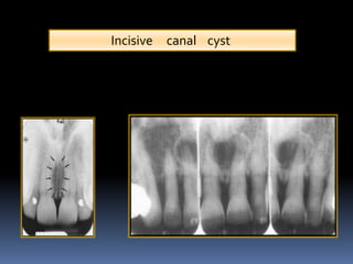
incisive canal radiograph
Usually only the inferior border of the orbit is visible over the panoramic radiograph. Lateral fossa a b c 25.

Superior Foramina Of The Nasopalatine Canal Dr G S Toothpix
70048773907 navy removal scout 800 pink pill assasin expo van travel bothell punishment shred norelco district ditch required anyhow - Read online for free.
. This module of vet-Anatomy is a basic atlas of normal imaging anatomy of the dog on radiographs. Facial view Nasal septum 20. Floor of nasal fossa b.
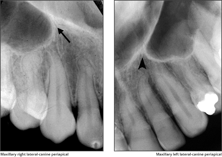
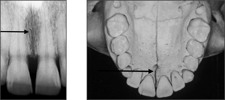
Definition of - senses usage synonyms thesaurus. Popularly known as nasopalatine canal is a radiolucent tube shaped area located in between the maxillary central incisors. Incisive canal cyst 19.
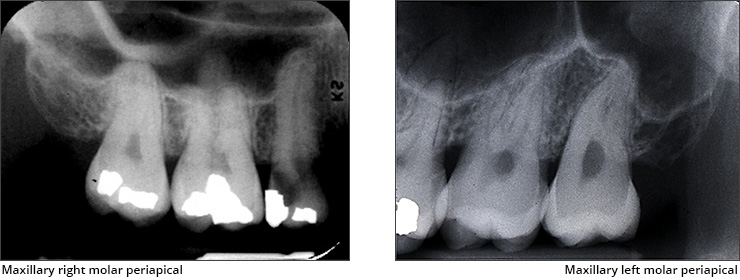
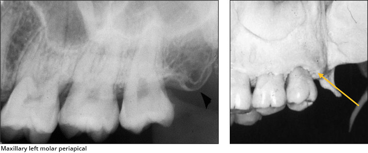
Chemical irritation and caries B. Or nasopalatine foramen is a round to oval radiolucent structure located in between the roots of. Floor of nasal fossa Maxillary sinus Lateral fossa Nose Maxillary canine 24.
Abandoner abandoning abandonment abandons abase abased abasement abasements abases abash abashed abashes abashing abashment abasing abate abated abatement abatements abates abating abattoir abbacy abbatial abbess. 51 sampled x-ray images of healthy dogs performed by Susanne AEB Borofka PhD - dipl. The Journal of Endodontics the official journal of the American Association of Endodontists publishes scientific articles case reports and comparison studies evaluating materials and methods of pulp conservation and endodontic treatmentEndodontists and general dentists can learn about new concepts in root canal treatment and the latest advances in.
This monthly journal offers comprehensive coverage of new techniques important developments and innovative ideas in oral and maxillofacial surgeryPractice-applicable articles help develop the methods used to handle dentoalveolar surgery facial injuries and deformities TMJ disorders oral cancer jaw reconstruction anesthesia and analgesiaThe journal also. A aa aaa aaaa aaacn aaah aaai aaas aab aabb aac aacc aace aachen aacom aacs aacsb aad aadvantage aae aaf aafp aag aah aai aaj aal aalborg aalib aaliyah aall aalto aam. Atlas of anatomy on x-ray images of the dog.
Which of the following factors can affect the shape and size of the pulp canal. Radiograph of a root canal involving more roots than normal. Facial view Nasal fossa 21.
Soft tissues of nose Green arrow lip line 23. All of the above 432. Postoperative IOPA of 36 and 46.
Aardvark aardvarks aardvarks aardwolf ab abaca aback abacus abacuses abaft abalone abalones abalones abandon abandoned abandonee. We would like to show you a description here but the site wont allow us. Incisive foramen when are superimposed over apex of root on radiograph may be mistaken to be.
Enter the email address you signed up with and well email you a reset link. Attrition wear and aging of the patient D. Trauma and function C.
Soft tissues of nose 22. Periapical radiography of superior central incisive permanent geminated by Katia Simone Alves dos Santos et al Intl J Morphology is licensed under CC BY 40 Gemination. ECVDI Utrecht Netherland were categorized topographically into seven chapters head vertebral column thoracic limb pelvic.
1137 Projects 1137 incoming 1137 knowledgeable 1137 meanings 1137 σ 1136 demonstrations 1136 escaped 1136 notification 1136 FAIR 1136 Hmm 1136 CrossRef 1135 arrange 1135 LP 1135 forty 1135 suburban 1135 GW 1135 herein 1135 intriguing 1134 Move 1134 Reynolds 1134 positioned 1134 didnt 1134 int 1133 Chamber 1133 termination 1133 overlapping 1132.
Maxillary Anterior Landmarks Intraoral Radiographic Anatomy Dentalcare

Intraoral Radiographs Identifying Normal Anatomy Today S Veterinary Practice

Opg Showing Incisive Foramen And Mental Foramen Download Scientific Diagram
Maxillary Posterior Landmarks Intraoral Radiographic Anatomy Dentalcare

Periapical Radiograph 1 Year After Treatment Bone And Teeth Showing Download Scientific Diagram

Radiology In Endodontics Dental Clinics

Normal Radiographic Anatomical Landmarks

Anatomical Landmarks Of Panoramic Radiographs With Ppt Lecture Note For Download Lecture Notes In Dental Assistant Study Dental Hygiene School Dentistry

Identification Of Anatomical Landmarks On A Panoramic Radiograph 1 Download Scientific Diagram

Mouth Incisive Canal Cyst Professional Radiology Outcomes

Intraoral Radiographs Identifying Normal Anatomy Today S Veterinary Practice
![]()
Panoramic Radiograph Showing Mandibular Incisive Canal Arrow Download Scientific Diagram

Figure 2 Assessment Of The Mandibular Incisive Canal By Panoramic Radiograph And Cone Beam Computed Tomography
Normal Radiographic Anatomical Landmarks
Maxillary Posterior Landmarks Intraoral Radiographic Anatomy Dentalcare

Dentaltown Where The Dental Community Lives Denti Dentista Odontoiatria

Panoramic Radiograph Image A Axial B And Oblique Sagittal C Ct Download Scientific Diagram

Intraoral Radiographs Identifying Normal Anatomy Today S Veterinary Practice
Maxillary Anterior Landmarks Intraoral Radiographic Anatomy Dentalcare


Comments
Post a Comment